Basic Science
Poster Session 3
(806) Validation of the fetal lamb model of complex gastroschisis

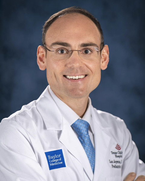
Luc Joyeux, MD, PhD
Texas Children's Hospital, Baylor College of Medicine
Houston, TX, United States- DB
David Basurto, MD, PhD
Academic Department of Development and Regeneration, Cluster Woman and Child, Biomedical Sciences
KU Leuven
Leuven, West-Vlaanderen, Belgium - KW
Kanokwaroon Watananirun, MD
My FetUZ Department of Development and Regeneration, Cluster Woman and Child, Biomedical Sciences
KU Leuven
Leuven, West-Vlaanderen, Belgium - TB
Tom Bleeser, MD, PhD
KU Leuven
Leuven, West-Vlaanderen, Belgium - IV
Ignacio J. Valenzuela, MD, PhD
KU Leuven
Leuven, West-Vlaanderen, Belgium - TA
Tomohiro Arai, MD
KU Leuven
Leuven, West-Vlaanderen, Belgium - ID
Inge Depoortere, PhD
KU Leuven
Leuven, West-Vlaanderen, Belgium - AA
Alison Accarie, PhD
KU Leuven
Leuven, West-Vlaanderen, Belgium - TV
Tim Vanuystel, MD, PhD
KU Leuven
Leuven, West-Vlaanderen, Belgium - MA
Michael Aertsen, MD
KU Leuven
Leuven, West-Vlaanderen, Belgium - AM
Alex Menys, PhD
Motilent
London, England, United Kingdom - PD
Paolo De Coppi, MD, PhD
Great Ormond Street Hospital, NHS
London, England, United Kingdom - LH
Larry H. Hollier, MD
Texas Children's Hospital, Baylor College of Medicine
Houston, TX, United States 
Michael A. Belfort, MD, PhD (he/him/his)
Professor, Chairman
Texas Children's Hospital
Houston, TX, United States
Jan Deprest, FRCOG
University Hospitals Leuven
Leuven, Vlaams-Brabant, Belgium- SK
Sundeep Keswani, MD
Texas Children's Hospital, Baylor College of Medicine
Houston, TX, United States
Primary & Presenting Author(s)
Coauthor(s)
Despite perceived favorable outcomes, complex Gastroschisis (cGS) has a significant morbidity such as sepsis, necrotizing enterocolitis, short bowel syndrome and bowel obstruction. A realistic animal model is lacking to enable further investigation of the pathogenesis of bowel dysfunction and development of novel prenatal therapies. We aimed to develop and validate a fetal lamb model of cGS.
Study Design:
In fetal lambs at 75 days’ gestation, a 1cm abdominal incision was made, a 1cm diameter rigid silicone constrictive ring was fixed to the abdominal wall and the small bowel was exteriorized through the standardized defect. At term (145d), lambs were delivered and compared to normal control lambs. Outcomes were small bowel gross examination, in vivo motility (primary) and structure on MRI, ex vivo contractility and permeability, and histological analysis.
Results:
5 of the 11 (45%) fetal lambs who underwent cGS induction survived to term. Compared to normal lambs (n=6), all lambs displayed a macroscopic phenotype of cGS (n=5): the small bowel and most of the large bowel were eviscerated, distended, thick and inflamed; there was small bowel obstruction with stenosis, atresia, necrosis and/or perforation (Fig.1A). On MRI, cGS lambs showed significantly decreased small bowel motility (motility GI-Quant score) and increased proximal bowel obstruction (motility contrast score), intra-abdominal bowel dilation and wall thickness (Fig.1B). They also had a reduced bowel contractility with significant decrease in smooth muscle contractions (Fig.1C). There were trend toward increased bowel permeability and increased integrity (Fig.1D). Histological analysis confirmed the abnormal architecture including thickening of all layers especially of proximal jejunum smooth muscle, serosal vascular proliferation, inflammatory cellular infiltrates in the serosa and submucosa, and shorter mucosal villi and crypts (Fig.1E-G).
Conclusion:
The fetal lamb model of cGS accurately replicates at term the structural and functional phenotype observed in human neonates with complex GS.

